Cornea anisotropy imaging using hybrid Brillouin-Raman micro-spectroscopy

Competition name: Zhas galym 2022-2024, MSHE RK

Project coordinator: Kharmysov C.

Total amount financed: 19 000 000 tenge

Project aim

The goal of the project is to develop and validate the hybrid Brillouin-Raman micro-spectroscopy technique for simultaneous non-contact assessment of viscoelastic and chemical imaging of cornea and apply image analysis, and machine learning techniques to analyze biomechanical and chemical profiles of a diseased and healthy cornea.

Objectives

To research the project goal, the following objectives are set, which are broken down into three financial years (FY):

  1. FY 2022: Brillouin micro-spectroscopy: viscoelastic (including anisotropic) analysis of probed cornea tissues correlated with collagen orientation
  2. FY 2023: Raman micro-spectroscopy: chemical analysis of probed cornea tissues.
  3. FY 2024: Hybrid Brillouin-Raman single point micro-spectroscopy and automated scan mapping of mechano-chemical spectra from cornea tissues. Correlation of viscoelastic and chemical properties with the results obtained from separate Brillouin and Raman micro-spectroscopies. Automated machine learning-based algorithm for the analysis of mechano-chemical images of cornea.

Expected results

According to the results of the implementation of scientific and (or) scientific and technical projects for the entire period of the project, the postdoctoral student should receive the following minimum results:

Publication of research results in the field of natural sciences, engineering, and technology, medicine, and healthcare, agricultural and veterinary sciences:

– at least 2 (two) articles in journals from the first three quartiles by impact factor in the Web of Science database or having a CiteScore percentile in the Scopus database of at least 50.

Current results

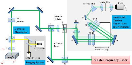
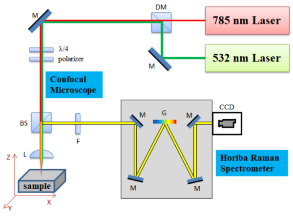
In the course of the research project, the team utilized Brillouin and Raman spectroscopy to examine the mechanical and chemical properties of biomaterials. This optical system enabled biosensing without causing any disturbance or harm to the samples. The outcomes obtained from the optical biosensor were compared with conventional methods, affirming its successful application in measuring molecular-level temperature variations. For the Brillouin site, we employed 6-pass Sandercock Tandem Fabry-Perot Interferometer (JRS instruments, Switzerland) coupled with a microscope in combination with Torus single longitudinal mode laser and imaging system (Figure 1) to measure Brillouin spectra from biomaterials. For the Raman measurements, we employed Horiba LabRAM HR Raman microscope. It is equipped with 532 and 633 nm laser wavelengths and provides Raman spectroscopic information with micro-scale confocal microscopy imaging capability. The schematic of the set-up used for the Raman Spectroscopy measurements is depicted in Figure 2.

Figure 1. Six-pass Tandem Fabry-Perot Interferometer coupled with microscope and laser

Figure 2. LabRAM HR Raman microscope.

2023

Q1 Journal Article

Mechano-Chemistry across Phase Transitions in Heated Albumin Protein Solutions

C Kharmyssov, K Sekerbayev, Z Nurekeyev, A Gaipov, ZN Utegulov

Polymers 15 (9), 2039

published


Research team members:

Kharmyssov, PhD, assistant professor, Department of Computer Engineering, Astana IT University. Scopus:57207846433; ID Web of Science: AAR-9491-2020; ORCID ID: 0000-0003-0683-0374; Publons   AAR-9491-2020. H-index: 2.

Scientific advisor:

Dzumatayeva Z.A. PhD, MD, professor. H index: 1. ORCID: 0000-0002-5383-3562